Courtesy of Dr. KW Chan and Dr. G. Chan, Department of Pathology, Queen Mary Hospital
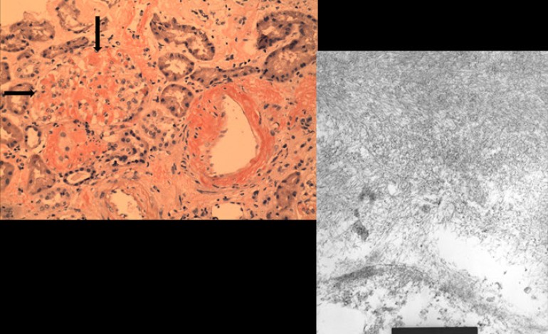
This is a renal biopsy from a patient with amyloidosis who presented with nephrotic-range proteinuria. The classical histopathological features include nodular glomerular lesions which are Congo Red positive (arrow). These nodular lesions will show apple-green birefringence under polarized light examination. Electron microscopy will reveal wavy amyloid fibrils with diameter ranging 5-25 nm.

